
INVITED Speakers

Confirmed Invited Speakers
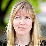 |
Carolina Wählby
LECTURE TITLE: Digital image analysis at microscopy resolution for better understanding of disease |
|
| Carolina Wählby is Professor in Quantitative Microscopy at the Dept. of Information Technology and Science for Life Laboratory, Uppsala University, Sweden. Her research is focused on algorithms for analysis of microscopy images with life science applications. She received a MSc in Molecular Biotechnology in 1998, a PhD in Digital Image Analysis in 2003, and carried out PostDoctoral research at the Dept. of Genetics and Pathology, all at Uppsala University. She joined the Broad Institute of Harvard and MIT in 2009 to work with the free and open source CellProfiler software, moving from cells to image large-scale image-based screening using model organisms such as C. elegans and zebrafish. Wählby returned to Sweden and became full professor in 2014, was elected ISAC scholar 2014, received the President’s innovation award from the Society of Biomolecular Imaging and Informatics in 2014, the Thuréus prize from The Royal Society of Sciences at Uppsala in 2015, and an ERC consolidator grant in 2015. Her current research includes methods development for large scale screening using cells and model organisms and digital pathology combined with in situ gene expression profiling. | ||
|
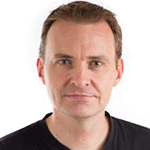
Bram van Ginneken
LECTURE TITLE: Computerized detection and quantification of lung diseases: assistant or replacement of human readers? |
|
| Bram van Ginneken is Professor of Functional Image Analysis at Radboud University Medical Center. Since 2010, he is co-chair of the Diagnostic Image Analysis Group within the Department of Radiology and Nuclear Medicine, together with Nico Karssemeijer. He also works for Fraunhofer MEVIS in Bremen, Germany, and is one of the founders of Thirona, a company that provides quantitative analysis of chest CT scans. Bram studied Physics at the Eindhoven University of Technology and at Utrecht University. In March 2001, he obtained his Ph.D. at the Image Sciences Institute (ISI) on Computer-Aided Diagnosis in Chest Radiography. From 2001 through 2009 he led the Computer-Aided Diagnosis group at ISI, where he still has an Associated Faculty position. He has (co-)authored over 150 publications in international journals. He is Associate Editor of IEEE Transactions on Medical Imaging and member of the Editorial Board of Medical Image Analysis. He is involved in organizing challenges in medical image analysis. | ||
 |
Fabrice De Chaumont
LECTURE TITLE: Tracking multiple mice for behavioral recognition |
|
| Fabrice de Chaumont has a PhD in Robotics (Design of robots navigating in
real time using stereo vision). He works on real time detection, automatic
behavior analysis and tracking. He is also the main leader of the open source software Icy
http://icy.bioimageanalysis.org. He is part of the team of BioImage
Analysis at the Institut Pasteur in Paris lead by Jean-Christophe
Olivo-Marin. The team aims at introducing innovative image processing and mathematical
approaches to biological imaging, and developing image analysis and
computer vision tools for the processing and quantification of
multi-channel temporal 3D sequences in biological microscopy. Main publications: Chenouard N, Smal I, de Chaumont F et al. Objective comparison of particle tracking methods. Nat Methods. 2014 de Chaumont F et al. Icy: an open bioimage informatics platform for extended reproducible research. Nat Methods. 2012 de Chaumont F, Coura RD, Serreau P, Cressant A, Chabout J, Granon S, Olivo-Marin JC. Computerized video analysis of social interactions Nat Methods. 2012 |
||
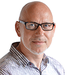 |
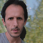
Jeroen van der Laak
LECTURE TITLE: Computer aided diagnosis will change the way we practice anatomical pathology |
|
| Jeroen van der Laak is Assistant Professor at the department of Pathology of the Radboud University Medical Center. He leads a research group in digital pathology, working in close collaboration with the diagnostic image analysis group (DIAG) of the Radboud UMC Radiology department. The main focus of his work is computer aided diagnosis in Pathology. His group develops and validates algorithms, often combined with specific laboratory techniques, to aid the diagnostic process. Jeroen has an MSc in computer science and acquired his PhD at the Radboud University in Nijmegen. He co-authored over 80 peer-reviewed publications, is member of the editorial board of the Journal of Pathology Informatics and organizer of sessions at the European Congress of Pathology and Pathology Visions. Jeroen acquired research grants from the European Union and the Dutch Cancer Society, among others. | ||
 |
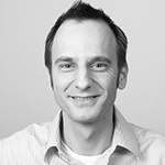
António Pereira
LECTURE TITLE: Cytoskeleton filament dynamics probed by speckled
photoswitching
|
|
| António Pereira received his BsC degree in Applied Physics followed by an MsC in Physics from the University of Minho under the supervision of Dr. Michael Belsley. He did his Ph.D. work on cell division at IBMC (University of Porto) under the supervision of Dr. Helder Maiato and Dr. Michael Belsley. His work focused on mathematical modeling and development of optical perturbation methods for studying mitotic spindle mechanics. Within the Chromosome Instability and Dynamics lab, he heads the imaging unit, which is devoted to the assembly and development of optical perturbation and imaging techniques, such as laser sub-micron surgery and inducible speckle imaging. | ||
 |
Jacco van Rheenen
LECTURE TITLE: High resolution intravital imaging of cancer plasticity
|
|
| Jacco van Rheenen was originally trained in a variety of imaging techniques during his PhD with Dr. Kees Jalink at the Netherlands Cancer Institute. He was among the first to optimize imaging and develop software to quantitatively measure FRET on confocal microscopes. During his PhD in the lab of Dr. Jalink and as postdoc in the lab of Dr. Sonnenberg (Netherlands Cancer Institute) he used several microscopy techniques to study lipid signaling in tumor cells. In order to broader his scales, he obtained a KWF fellowship to do a postdoc in the United States in the lab of Dr. John Condeelis. There he extended his imaging experience by imaging mammary tumors intravitally including two-photon microscopy and became an expert in the field of intravital FRET imaging. In 2008 he was appointed as group leader at the Hubrecht Institute, where he utilizes his imaging techniques to visualize processes that are required for the metastasis of mammary tumor cells in living animals. In 2009, he was awarded a VIDI grant and a research grant from the Dutch Cancer Society. In 2012, he was awarded a research grant from the Association for International Cancer Research (who have now rebranded to Worldwide Cancer Research), and in 2013 a research grant from Netherlands Organisation for Scientific Research. In 2013, he received the Stem Cells Young Investigator Award (see video below). In July 2014 he was appointed professor in Intravital Microscopy at the University Medical Center Utrecht. In 2015, he was awarded an ERC consolidator grant. |
||
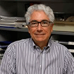 |
Daniel Navajas
LECTURE TITLE: Probing cell and tissue nanomechanics with Atomic Force
Microscopy |
|
| Daniel Navajas is Professor of Physiology (from 1989) at the Unit of
Biophysics and Bioengineering of the School of Medicine and Health Sciences
of the University of Barcelona (UB) and Group Leader (from 2007) at the
Institute for Bioengineering of Catalonia (IBEC). He is also Director of
Education (from 2010) for the Degree on Biomedical Engineering at UB. His
main research interest is the study of the mechanical behavior of the
respiratory system with a multiscale approach, ranging from cellular
nanomechanics to pulmonary pathophysiology with the goal of improving the
diagnosis and treatment of respiratory diseases. He has published more than
180 papers in journals indexed in the Journal Citation Reports. His publications received over 14,000 citations with an H-index of 40. |
||
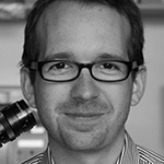 |
Benjamin Victor Ineichen
LECTURE TITLE: Can Nogo-A-antibodies boost neuro-rehabilitation in
(chronic) Multiple Sclerosis?
|
|
| Benjamin Victor Ineichen is a senior MD-PhD candidate in the laboratory of
Professor Martin Schwab at the University and ETH Zurich in Switzerland. Martin Schwab was the pioneer in isolating Nogo-A, one of the most potent nerve fiber growth inhibitors. His lab is world-renowned for its research on neuronal plasticity and the investigation of ways of how to boost neuronal repair of spinal cord injuries and strokes in order to achieve neuro-rehabilitation. Furthermore, his research group conducts a clinical trial testing the effects of anti-Nogo-A antibodies in spinal cord injury patients. Benjamin Victor Ineichen co-investigates with Martin Schwab the role of the inhibitory protein Nogo-A in Multiple Sclerosis (MS) and how Nogo-A-antibodies could be used to promote neuro-rehabilitation in MS. His group is mainly interested how myelin, the insulating sheaths of nerve fibers, can be restored to enable efficient nerve conduction again leading to overlying rehabilitation. He developed a new method to image repaired myelin sheaths on a large scale to screen for potential novel remyelinating drugs. Benjamin Victor Ineichen received his medical degree in 2012 at the University of Zurich and did his MD-thesis in neurometabolics at the University Hospital of Zurich under the supervision of Professor Michael Linnebank. In 2010, he completed a Neuroradiology Fellowship at the University of Pennsylvannia under Professor Elias Melhem. Additionally, he was a finalist at the „falling walls lab“ in Berlin 2015 (young innovator of the year) and he was awarded research grants from the Swiss Academy of Medical Sciences and from the Danish Desirée and Niels-Yde Foundation, dedicated to the treatment of Multiple Sclerosis. After his PhD, he will specialize in Neuroradiology. |
||
Address: Rua Alfredo Allen, 208 | 4200-135 Porto, Portugal | Phone: +351 220 408 800 | Email: events@i3s.up.pt
| ORGANIZED BY: | CO-ORGANIZED: | SUPPORT: | |||
 |
 |

 |
 |