Lab Sessions
Each lab session has a limited number of seats. Registration based of first-come, first-served.
A. Atomic Force Microscopy in the context of Bioimaging
Instructors: Manuela Brás, i3S-INEB; Susana Sousa, i3S-INEB
i3S - Instituto de Investigação e Inovação em Saúde
Rua Alfredo Allen, 208, 4200-393 Porto, Portugal
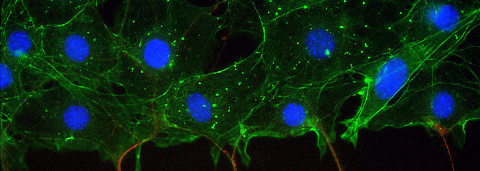
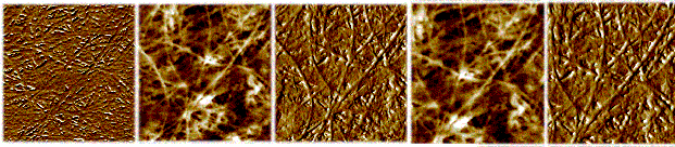
Atomic Force Microscopy (AFM) is a powerful technique to perform dynamic and biomechanical studies on different cell types. For that, one or more cantilevers, mounted at the end of a piezoelectric material on a chip moves across a selected area of the cell surface. Interatomic forces between the probe tip and the cell cause the cantilever to deflect as the cells’ surface topography changes. A laser light reflected from the back of the cantilever measures the deflection of the cantilever. The probe (that can be spherical or pyramidal shaped) can be used as a mechanical sensor to performed nanoindentation (nanomechanical) studies. Mechanobiology is focused on understanding the “mechanotransduction” of cells that is defined as the mechanisms by which cells convert mechanical stimulus into biochemical signals/ activity.
The mechanical properties (stiffness, Young’s modulus, adhesion, deformation and viscolelasticity) of cells are important for many cellular processes like cell migration, cell protrusion, cell division, cell morphology, in order to understand several mechanisms like some of those found at oncologic diseases. To observe precisely the region of interest on the sample, the AFM will be coupled on the bottom of an Inverted Fluorescence Microscope (IFM). In this lab session the participants will be introduced to the AFM/IFM techniques and the nanomechanical properties of fresh human tissues cells (colorectal tumor and adjacent tumor tissues) will be assessed.
B. Microscopy image processing and data extraction with ICY
Instructors: Jerome Mutterer, CNRS - Centre National de la Recherche Scientifique
Atmosfera M | Rua Júlio Dinis, nº 158/160, 5th floor | 4050-318 Porto, Portugal
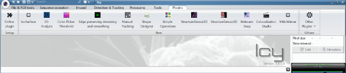
An image is worth a thousand... data points, and modern microscopy imaging techniques provides access to information rich images. Efficient data extraction from images requires however knowledge regarding different techniques, and the conditions of applicability and limitations of each one. Access to a basic "bag of tools" in image processing can be very useful to researchers interested in quantitative imaging.
This is a hands-on lab session, where the participants will learn and explore different image processing methodologies with relevance for microscopy. The well-established, and free, ICY software will be used to tackle case studies addressing common problems such as segmentation, morphometry and particle counting.
Address: Rua Alfredo Allen, 208 | 4200-135 Porto, Portugal | Phone: +351 226 074 900 | Email: events@i3s.up.pt
| ORGANIZED BY: | CO-ORGANIZED: | SUPPORT: | |||
 |

|
 |